EMG reported as gamma
Symptom. Broadband high-frequency power over temporal/frontal sites tied to jaw or neck.
Symptom
A “gamma” effect (conventionally above about 30 Hz) that is largest over temporal, lateral frontal or occipital electrodes, is broadband rather than peaked, and appears in conditions where the subject is more likely to be tense, talking, chewing, smiling or straining: effortful tasks, emotional stimuli, speech, movement preparation. Single-subject spectra show a raised high-frequency floor rather than a bump. Consumer and dry-electrode recordings show it more, because higher impedances and looser contact pick up more muscle.
Cause
Scalp muscles (temporalis, frontalis, masseter, the neck extensors) produce electrical activity that is large compared with cortical gamma, broadband from roughly 20 Hz upward with no single peak, and sits directly under the electrodes that “show the effect”. Its amplitude is modulated by anything that changes muscle tone — and many experimental manipulations do. The spectrum of EMG overlaps the whole gamma range, so a band-power measure cannot separate the two, and a condition difference in muscle tone becomes a condition difference in “gamma”.
Detect
- Topography: cortical gamma from a focal source is rarely largest at the edge of the montage; EMG is. Look at T7/T8, F7/F8, FT sites and the occipital rim.
- Spectral shape: EMG raises the floor across a wide range with no peak; a genuine oscillation has a peak above the aperiodic background (L1.7, L4.6).
- Time course: EMG turns on and off with muscle events (swallows, jaw movement, blinks with a squint) rather than with the stimulus at a fixed latency.
- Condition confound: ask whether one condition plausibly involves more tension, speech or facial expression than the other.
- In ICA space, EMG components have a spatially restricted rim topography and a flat, high-frequency spectrum (L2.6).
Fix
- Instruct and monitor: relaxed jaw, no talking, breaks; record a facial EMG channel where high frequencies matter.
- Clean before measuring: ICA with muscle-component removal, or rejection of segments with high-frequency power above a threshold (L2.5, L2.6); state what was removed.
- Report a peak, not a band: use spectral parameterization to show a gamma peak above the aperiodic background before calling it gamma.
- Prefer measures that are robust to broadband noise, such as inter-trial phase consistency at a specific frequency (L4.5), and check that the effect survives on the cleaned data.
- Be conservative in low-density and consumer recordings, whose hardware bandwidth may not even reach the gamma band (see pf-hardware-bandwidth-ceiling).
Example
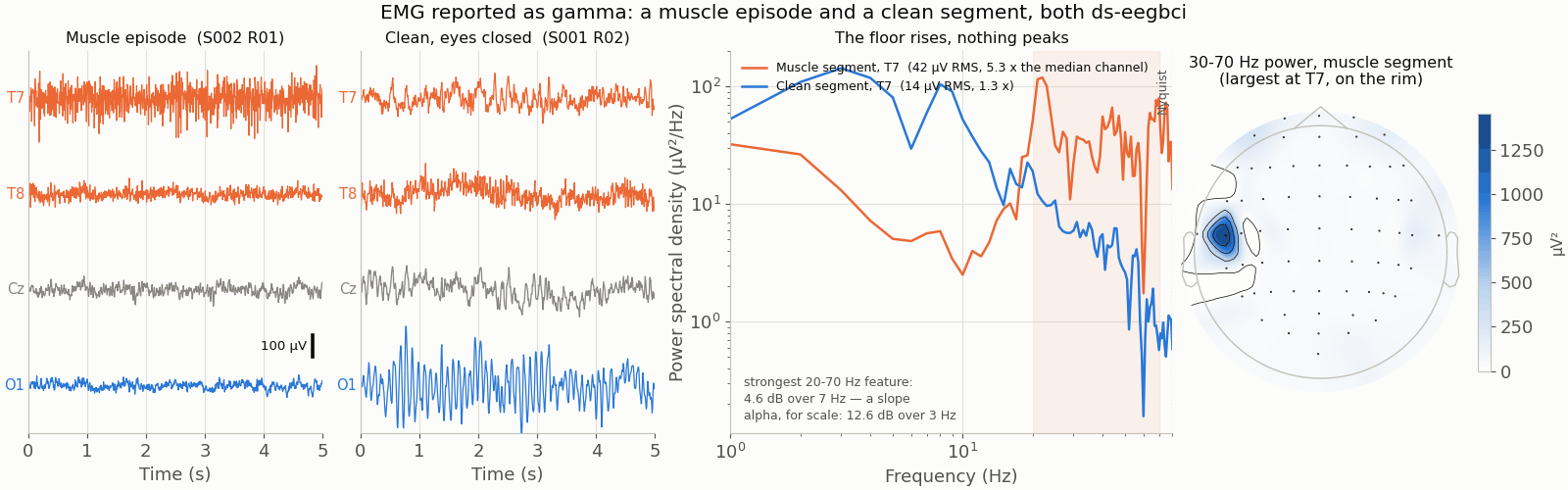
The figure, generated by data/scripts/make_figures_p4.py: a 5-s segment with a jaw-clench episode over T7/T8, its PSD showing a raised high-frequency floor with no peak, and the corresponding topography with power concentrated at the lateral rim — beside a segment of clean eyes-closed EEG whose PSD shows the alpha peak and a low, falling high-frequency floor.